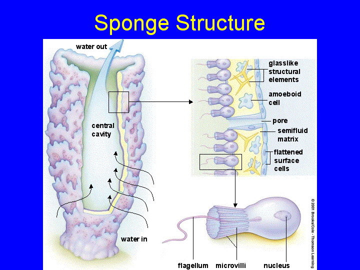


Whereas pinacocytes line the outside of the sponge, choanocytes tend to line certain inner portions of the sponge body that surround the mesohyl. In other sponges, ostia are formed by folds in the body wall of the sponge.Ĭhoanocytes (“collar cells”) are present at various locations, depending on the type of sponge, but they always line the inner portions of some space through which water flows (the spongocoel in simple sponges, canals within the body wall in more complex sponges, and chambers scattered throughout the body in the most complex sponges). In some sponges, ostia are formed by porocytes, single tube-shaped cells that act as valves to regulate the flow of water into the spongocoel. In addition to the osculum, sponges have multiple pores called ostia on their bodies that allow water to enter the sponge. The gel-like consistency of mesohyl acts like an endoskeleton and maintains the tubular morphology of sponges. Mesohyl is an extracellular matrix consisting of a collagen-like gel with suspended cells that perform various functions. Pinacocytes, which are epithelial-like cells, form the outermost layer of sponges and enclose a jelly-like substance called mesohyl. While sponges (excluding the hexactinellids) do not exhibit tissue-layer organization, they do have different cell types that perform distinct functions.

However, sponges exhibit a range of diversity in body forms, including variations in the size of the spongocoel, the number of osculi, and where the cells that filter food from the water are located. Water entering the spongocoel is extruded via a large common opening called the osculum. Water can enter into the spongocoel from numerous pores in the body wall. Trending Questions What happens when you have sun stroke? What is the limit beyond which stars suffer internal collapse called? What does the 23.The morphology of the simplest sponges takes the shape of a cylinder with a large central cavity, the spongocoel, occupying the inside of the cylinder.


 0 kommentar(er)
